JCRB1032 OZ
Cell information
Important Notice(s)On the agreement for distribution of JHH series cell lines, NOZ and OZ
Cell type:general cells (View Pricing Information)
| JCRB No. | JCRB1032 | Cell Name | OZ |
|---|---|---|---|
| Profile | Human bile duct cell line established from ascites of the tumor patient who had differentiated adenocarcinoma. | Other Name | |
| Animal | human | Strain | |
| Genus | Homo | Species | sapiens |
| Sex | M | Age | 71 |
| Identity | available | Tissue for Primary Cancer | liver, gallbladder |
| Case history | differentiated adenocarcinoma papilla and poorly differentiated adenocarcinoma. CEA 8.0 ng/ml. | Metastasis | Yes |
| Tissue Metastasized | ascites | Genetics | No. of chromosome, 51. Td=48 hrs. |
| Life Span | infinite | Crisis PDL | |
| Morphology | epithelial-like | Character | Cells were established from ascites of the patient whose bile duct was emphraxed by mucinous materials produced by adenocarcinoma cells. Cells produce CEA, BMG. High levels of P1-P, r-GTP, LDH were observed in cells. PAS (+). Transplantalbe to nude mice. |
| Classify | tumor | Established by | Honma,S. & Nagamori,S. |
| Registered by | Nagamori,S. | Regulation for Distribution | Require negotiation and agreement from Dr.Nagamori before shipping. |
| Comment | Cell Bank will handle to get the approval but the depositor may contact to the requested person directly. (r-GPT = gamma GPT) | Year | 2001 |
| Medium | Williams'E medium with 10% fetal calf serum. | Methods for Passages | Cells are harvested after treatment with 0.25% trypsin and 0.02% EDTA. |
| Cell Number on Passage | Race | Japanese | |
| CO2 Conc. | 5 % | Tissue Sampling | ascites |
| Tissue Type |
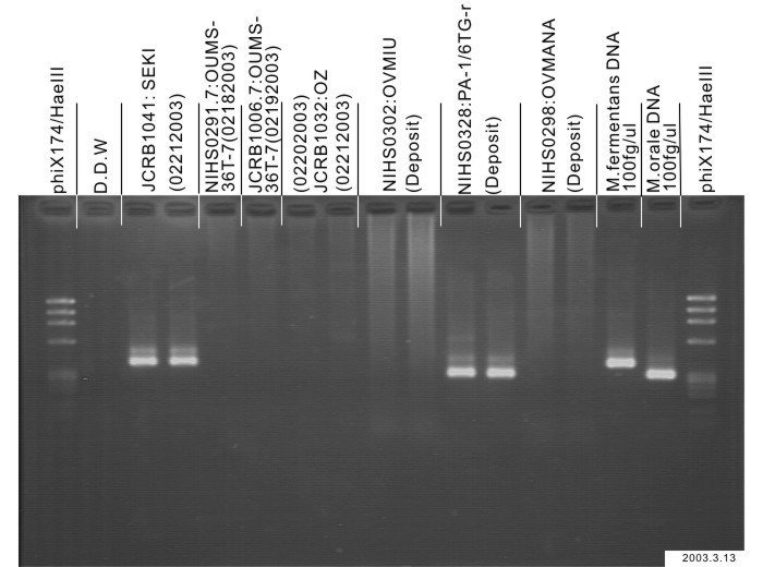
| Detection of virus genome fragment by Real-time PCR | |||||||||
|---|---|---|---|---|---|---|---|---|---|
| Detected DNA Virus | tested | Detected RNA Virus | tested | ||||||
| CMV | - | parvoB19 | - | HCV | - | HTLV-1 | - | ||
| EBV | - | HBV | - | HIV-1 | - | HTLV-2 | - | ||
| HHV6 | - | HTLV-1 | - | HIV-2 | - | HAV | - | ||
| HHV7 | - | HTLV-2 | - |
-/negative. +/positive. nt/not tested. (positive (+) does not immediately mean the production of infectious viral particles.) |
|||||
| BKV | - | HIV-1 | - | ||||||
| JCV | - | HIV-2 | - | ||||||
| ADV | - | HPV18 | - | ||||||
| Notes | |||||||||
| Reference | |
|---|---|
| Pubmed id:2889643 | Human bile duct carcinoma cell line producing abundant mucin in vitro. Homma S,Nagamori S,Fujise K,Yamazaki K,Hasumura S,Sujino H,Matsuura T,Shimizu K,Kameda H,Takaki K Gastroenterol Jpn. 1987 Aug;22(4):474-9 |
| Research results by users. Click! | |
|---|---|
| Pubmed id:23084587 |
Overexpression of epithelial-mesenchymal transition-related markers according to cell dedifferentiation: clinical implications as an independent predictor of poor prognosis in cholangiocarcinoma. Ryu HS,Chung JH,Lee K,Shin E,Jing J,Choe G,Kim H,Xu X,Lee HE,Kim DG,Lee H,Jang JJ Hum Pathol. 2012 Dec;43(12):2360-70 |
| Pubmed id:22639408 |
Twisted gastrulation expression in cholangiocellular and hepatocellular carcinoma. Johnston J,Al-Bahrani R,Abuetabh Y,Chiu B,Forsman CL,Nagamori S,Leng R,Petryk A,Sergi C J Clin Pathol. 2012 Oct;65(10):945-8 |
| Pubmed id:22457476 |
Therapeutic potential of amanitin-conjugated anti-epithelial cell adhesion molecule monoclonal antibody against pancreatic carcinoma. Moldenhauer G,Salnikov AV,Lüttgau S,Herr I,Anderl J,Faulstich H J Natl Cancer Inst. 2012 Apr 18;104(8):622-34 |
| Pubmed id:22309595 |
Fibroblast growth factor 19 expression correlates with tumor progression and poorer prognosis of hepatocellular carcinoma. Miura S,Mitsuhashi N,Shimizu H,Kimura F,Yoshidome H,Otsuka M,Kato A,Shida T,Okamura D,Miyazaki M BMC Cancer. 2012 Feb 6;12():56 |
| Pubmed id:22075503 |
Expression of E-cadherin and β-catenin in two cholangiocarcinoma cell lines (OZ and HuCCT1) with different degree of invasiveness of the primary tumor. Abuetabh Y,Persad S,Nagamori S,Huggins J,Al-Bahrani R,Sergi C Ann Clin Lab Sci. 2011 Summer;41(3):217-23 |
| Pubmed id:20922056 |
Mesothelin as a potential therapeutic target in human cholangiocarcinoma. Yu L,Feng M,Kim H,Phung Y,Kleiner DE,Gores GJ,Qian M,Wang XW,Ho M J Cancer. 2010 Oct 1;1():141-9 |
| Pubmed id:20565817 |
Expression of growth factor receptors and targeting of EGFR in cholangiocarcinoma cell lines. Xu L,Hausmann M,Dietmaier W,Kellermeier S,Pesch T,Stieber-Gunckel M,Lippert E,Klebl F,Rogler G BMC Cancer. 2010 Jun 18;10():302 |
| Pubmed id:20044606 |
Infrequent amplification of JUN in hepatocellular carcinoma. Endo M,Yasui K,Nakajima T,Gen Y,Tsuji K,Dohi O,Zen K,Mitsuyoshi H,Minami M,Itoh Y,Taniwaki M,Tanaka S,Arii S,Okanoue T,Yoshikawa T Anticancer Res. 2009 Dec;29(12):4989-94 |
| Pubmed id:19319137 |
Vandetanib (ZD6474), an inhibitor of VEGFR and EGFR signalling, as a novel molecular-targeted therapy against cholangiocarcinoma. Yoshikawa D,Ojima H,Kokubu A,Ochiya T,Kasai S,Hirohashi S,Shibata T Br J Cancer. 2009 Apr 21;100(8):1257-66 |
| Pubmed id:19453019 |
Rapamycin inhibits growth of cholangiocarcinoma cells. Okada T,Sawada T,Kubota K Hepatogastroenterology. 2009 Jan-Feb;56(89):6-10 |
| Pubmed id:19383345 |
Exon 2 deletion splice variant of gamma-glutamyl carboxylase causes des-gamma-carboxy prothrombin production in hepatocellular carcinoma cell lines. Ueda N,Shiraha H,Fujikawa T,Takaoka N,Nakanishi Y,Suzuki M,Matsuo N,Tanaka S,Nishina S,Uemura M,Takaki A,Shiratori Y,Yamamoto K Mol Oncol. 2008 Oct;2(3):241-9 |
| Pubmed id:18442209 |
Effect of histone deacetylase inhibitor on proliferation of biliary tract cancer cell lines. Xu LN,Wang X,Zou SQ World J Gastroenterol. 2008 Apr 28;14(16):2578-81 |
| Pubmed id:15492814 |
Anti-endostatin monoclonal antibody enhances growth of human hepatocellular carcinoma cells by inhibiting activity of endostatin secreted by the transplanted cells in nude mice. Tsuboi S,Nouso K,Tomono Y,Nagamori S,Naito I,Watanabe T,Manabe K,Ohmori H,Nakatsukasa H,Miyazaki M,Higashi T,Shiratori Y,Namba M,Ninomiya Y Int J Oncol. 2004 Nov;25(5):1267-71 |
| Pubmed id:12654561 |
Development of a therapeutic adenoviral vector for cholangiocarcinoma combining tumor-restricted gene expression and infectivity enhancement. Nagi P,Vickers SM,Davydova J,Adachi Y,Takayama K,Barker S,Krasnykh V,Curiel DT,Yamamoto M J Gastrointest Surg. 2003 Mar-Apr;7(3):364-71 |
| Pubmed id:9446792 |
Cyclin E overexpression responsible for growth of human hepatic tumors with p21WAF1/CIP1/SDI1. Tsuji T,Miyazaki M,Fushimi K,Mihara K,Inoue Y,Ohashi R,Ohtsubo M,Hamazaki K,Furusako S,Namba M Biochem Biophys Res Commun. 1998 Jan 14;242(2):317-21 |
| Pubmed id:9135018 |
Tamoxifen-mediated growth inhibition of human cholangiocarcinoma. Sampson LK,Vickers SM,Ying W,Phillips JO Cancer Res. 1997 May 1;57(9):1743-9 |
| Pubmed id:8835345 |
Persistence of hepatitis C virus RNA in established human hepatocellular carcinoma cell lines. Tsuboi S,Nagamori S,Miyazaki M,Mihara K,Fukaya K,Teruya K,Kosaka T,Tsuji T,Namba M J Med Virol. 1996 Feb;48(2):133-40 |
| Pubmed id:1701409 |
Integration of hepatitis B virus DNA into cells of six established human hepatocellular carcinoma cell lines. Fujise K,Nagamori S,Hasumura S,Homma S,Sujino H,Matsuura T,Shimizu K,Niiya M,Kameda H,Fujita K Hepatogastroenterology. 1990 Oct;37(5):457-60 |
| Pubmed id:2889643 |
Human bile duct carcinoma cell line producing abundant mucin in vitro. Homma S,Nagamori S,Fujise K,Yamazaki K,Hasumura S,Sujino H,Matsuura T,Shimizu K,Kameda H,Takaki K Gastroenterol Jpn. 1987 Aug;22(4):474-9 |
| Pubmed id:3005723 |
[Effects of hyperthermia on the proliferation and viability of cultured human hepatobiliary carcinoma cells]. Homma S,Nagamori S,Fujise K,Sujino H,Matsuura T,Simizu K,Kameda H Nihon Shokakibyo Gakkai Zasshi. 1985 Sep;82(9):2043-8 |
| Pubmed id:none |
高分化型ヒト不死化肝細胞株(OUMS‐29)と肝癌細胞株(JHH‐1)でのエンドスタチン分泌の相違 友野靖子 安藤香織 内藤一郎 佐渡義一 坪井壮 永森静志 宮崎正博 沖垣達 二宮善文 重井医学年報 24, 7-14 (2003) |
| Pubmed id:none |
樹立ヒト肝癌株細胞中のB型肝炎ウイルスゲノム 藤瀬 清隆, 藤多 和信, 永森 静志, 蓮村 哲, 本間 定, 筋野 甫, 松浦 知和, 清水 恵一郎, 新谷 稔, 大野 典也, 亀田 治男 肝臓 29(5), 697-698 (1988) |
| Pubmed id:none |
ヒト肝胆道系癌培養細胞の生存と増殖に及ぼす温熱の効果 本間 定, 永森 静志, 藤瀬 清隆, 筋野 甫, 松浦 知和, 清水 恵一郎, 亀田 治男 肝臓 24(5), 569 (1983) |
| Images |
|---|
                    |
| Movies |
|---|
|
LOT Information
| Cell No. | JCRB1032 | Cell Name | |
|---|---|---|---|
| LOT No. | 05222012 | Lot Specification | distribution |
| Medium | Williams' E medium with 10% fetal bovine serum (FBS, GIBCO Cat. # 10099) | Temperature | 37 C |
| Cell Density at Seeding | 1 - 3 x 10^5 cells/mL | Methods for Passages | Cells were harvested after treatment with 0.25% trypsin and 0.02% EDTA. |
| Doubling Time | 3 - 4 days | Cell Number in Vial (cells/1ml) | 2.0 x 10^6 |
| Viability at cell freezing (%) | 93.0 | Antibiotics Used | free |
| Passage Number | Unknown (16 at bank) | PDL | |
| Sterility: MYCOPLASMA | - | Sterility: BACTERIA | - |
| Sterility: FUNGI | - | Isozyme Analysis | Confirmed as human by NP, G6PD (type B), MD. |
| Chromosome Mode | Chromosome Information | ||
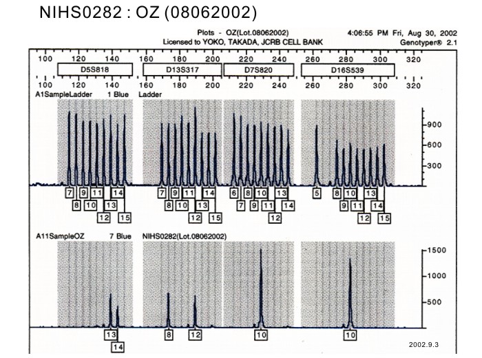
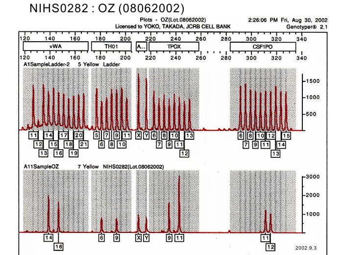
| Surface Antigen | DNA Profile (STR) | ||
| Adhesion | Yes | Exoteric Gene | |
| Medium for Freezing | 10% DMSO, 20% FBS - Williams' E | CO2 Conc. | 5% |
| Viability immediately after thawing (%) | Additional information |
| Cell No. | JCRB1032 | Cell Name | |
|---|---|---|---|
| LOT No. | 03192007 | Lot Specification | distribution |
| Medium | William's E medium with 10% fetal bovine serum (FBS lot; GIBCO 484375) | Temperature | 37 C |
| Cell Density at Seeding | approx. 1 x 10^5 cells/ml | Methods for Passages | 0.25% trypsin and 0.02% EDTA, passage once a week. Cells attached hard to dishes and trypsinization at 37 C for 20-30 minutes required. |
| Doubling Time | 80 hrs | Cell Number in Vial (cells/1ml) | 1.8 x 10^6 |
| Viability at cell freezing (%) | 98.3 | Antibiotics Used | free |
| Passage Number | Unknown (9 at bank) | PDL | |
| Sterility: MYCOPLASMA | - | Sterility: BACTERIA | - |
| Sterility: FUNGI | - | Isozyme Analysis | Confirmed as human by NP and G6PD, LD, MD. |
| Chromosome Mode | Chromosome Information | ||
| Surface Antigen | DNA Profile (STR) | ||
| Adhesion | Yes | Exoteric Gene | |
| Medium for Freezing | 10% DMSO, 20% FBS - William's E medium | CO2 Conc. | 5% |
| Viability immediately after thawing (%) | Additional information |
| Cell No. | JCRB1032 | Cell Name | OZ |
|---|---|---|---|
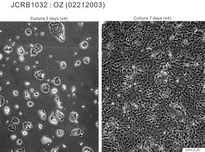
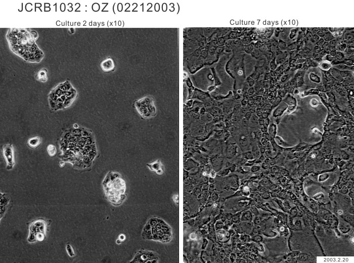
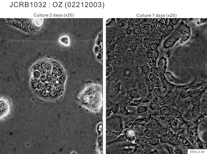
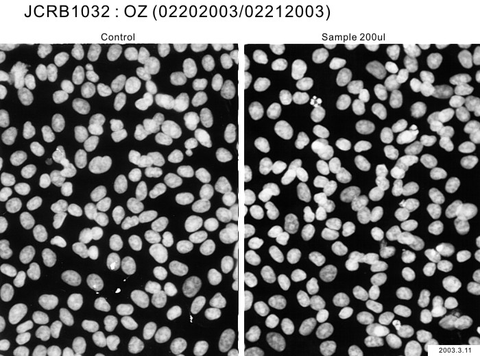
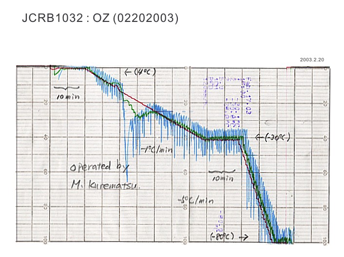
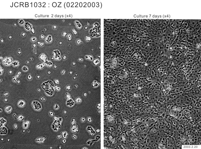
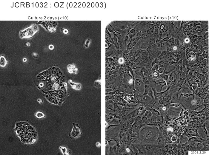
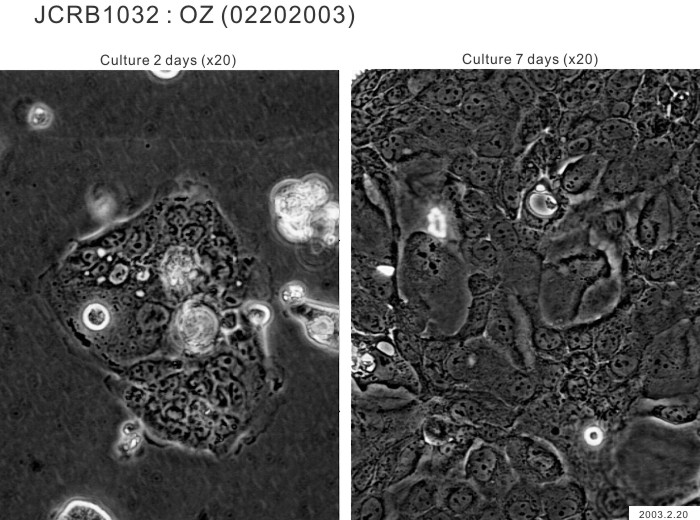
| LOT No. | 02212003 | Lot Specification | distribution |
| Medium | Williams'E medium with 10% fetal calf serum(Intergen RB52305). | Temperature | 37 C |
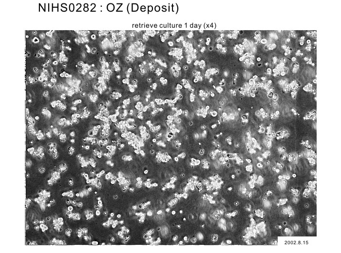
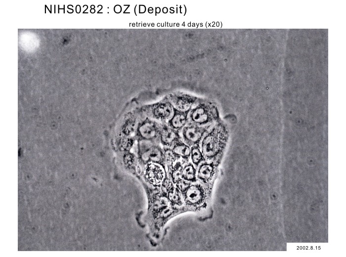
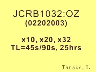
| Cell Density at Seeding | 1.2 x 10^4 cells/sq.cm. | Methods for Passages | Cells were harvested after treatment with 0.2% trypsin. Medium change twice a week and subculture every 10 days.. |
| Doubling Time | NT | Cell Number in Vial (cells/1ml) | 1.4 x 10^6 |
| Viability at cell freezing (%) | 96.1 | Antibiotics Used | free |
| Passage Number | P6* | PDL | NT |
| Sterility: MYCOPLASMA | - | Sterility: BACTERIA | - |
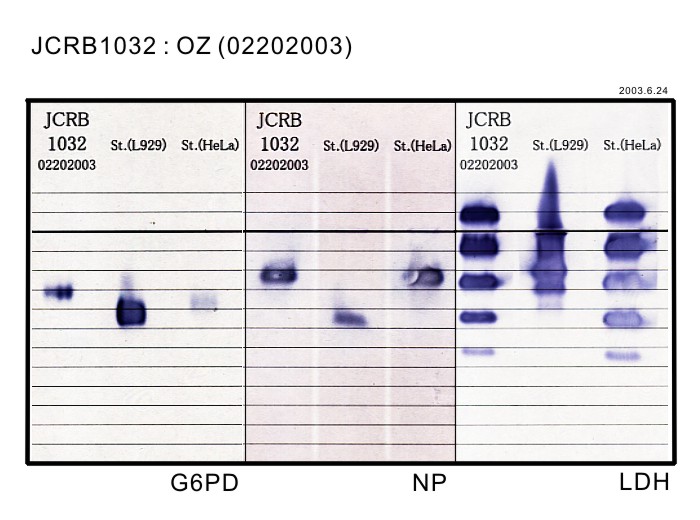
| Sterility: FUNGI | - | Isozyme Analysis | G6PD(not shown), NP, LDH examined. Human. |
| Chromosome Mode | NT | Chromosome Information | NT |
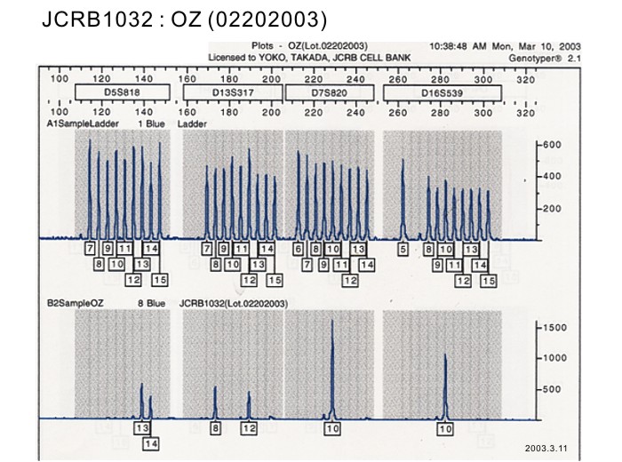
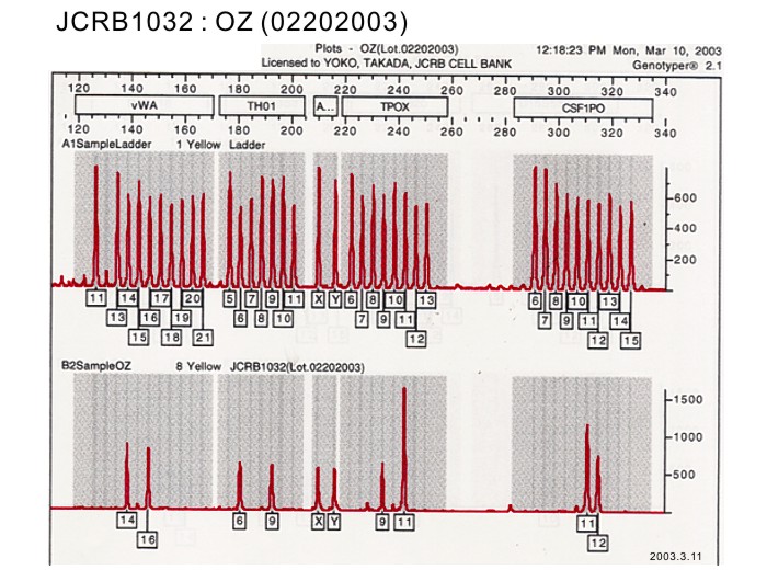
| Surface Antigen | NT | DNA Profile (STR) | |
| Adhesion | Yes | Exoteric Gene | NT |
| Medium for Freezing | Culture medium with 5% DMSO. | CO2 Conc. | 5 % |
| Viability immediately after thawing (%) | NT | Additional information |
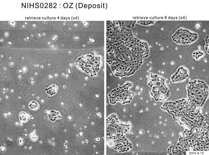
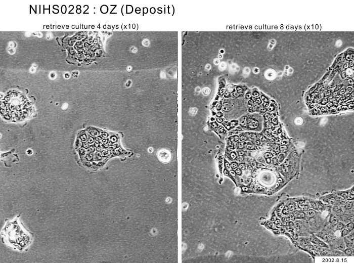
| Images |
|---|
|
|
| Cell No. | JCRB1032 | Cell Name | OZ |
|---|---|---|---|
| LOT No. | 12092020 | Lot Specification | distribution |
| Medium | Williams' E medium with 10% fetal bovine serum (FBS, Sigma Cat. # 172012, Lot 12J396) | Temperature | 37 C |
| Cell Density at Seeding | 3.6 - 5.4 x 10^4 cells/mL | Methods for Passages | Cells were harvested after treatment with 0.2% trypsin. |
| Doubling Time | approx. 44 hrs. | Cell Number in Vial (cells/1ml) | 6.1 x 10^5 |
| Viability at cell freezing (%) | 96 | Antibiotics Used | free |
| Passage Number | Unknown (10 at bank) | PDL | |
| Sterility: MYCOPLASMA | - | Sterility: BACTERIA | - |
| Sterility: FUNGI | - | Isozyme Analysis | |
| Chromosome Mode | Chromosome Information | ||
| Surface Antigen | DNA Profile (STR) | ||
| Adhesion | Yes | Exoteric Gene | |
| Medium for Freezing | 10% DMSO, 20% FBS - Williams' E | CO2 Conc. | 5% |
| Viability immediately after thawing (%) | 76 | Additional information |