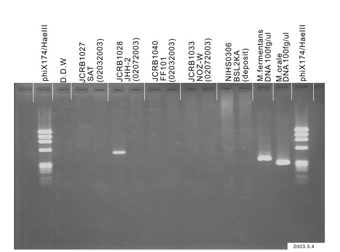
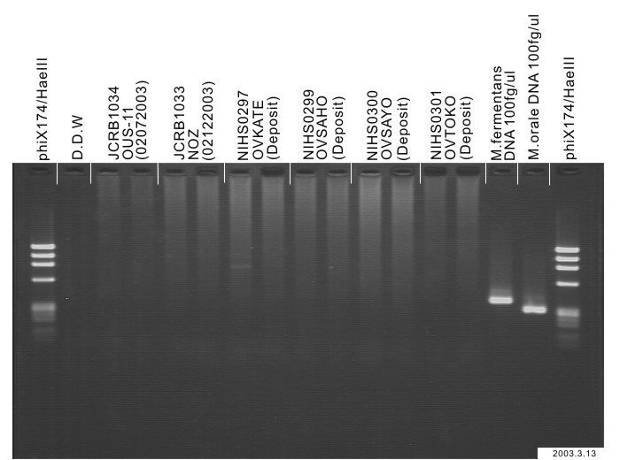
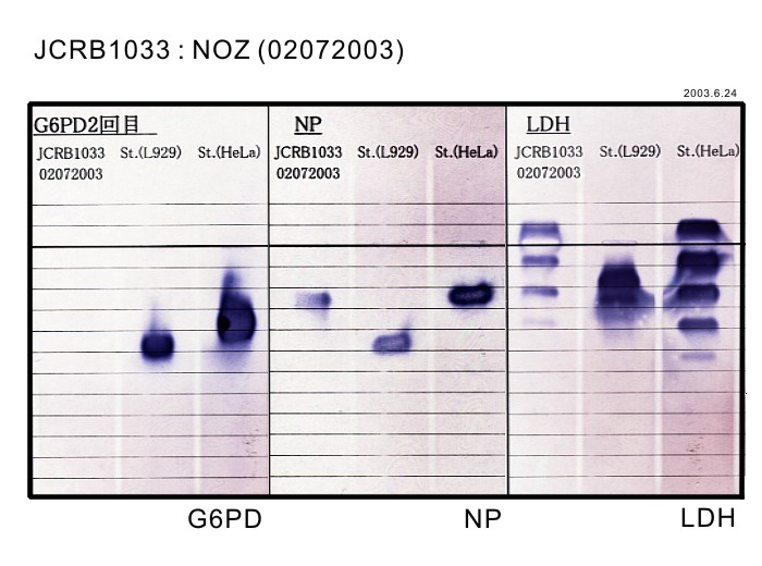
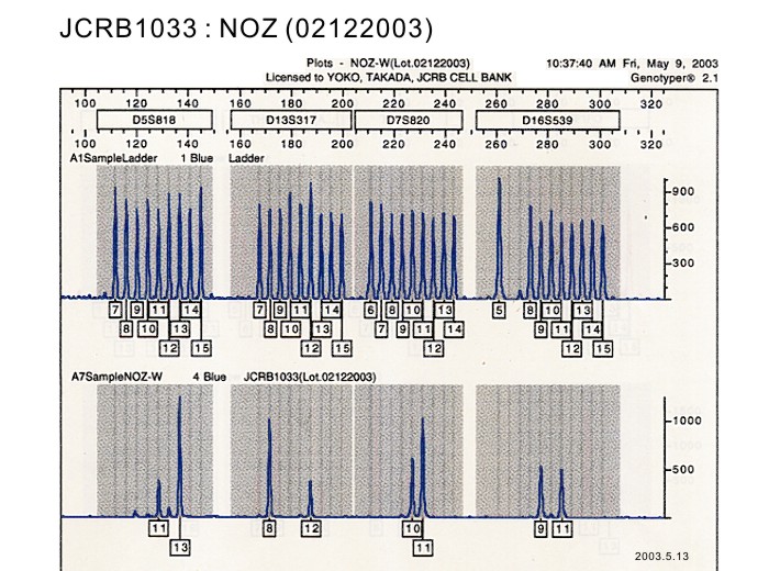
JCRB1033 NOZ
細胞情報
Important Notice(s)On the agreement for distribution of JHH series cell lines, NOZ and OZ
細胞種類:一般細胞 (細胞分譲手数料はこちら)
| 細胞番号(JCRB) | JCRB1033 | 細胞名 | NOZ |
|---|---|---|---|
| 生物種(日本語) | ヒト | 組織名(日本語) | 肝臓, 胆嚢 |
| コメント(日本語) | 胆嚢がん, 中分化型管状腺がん | プロフィール | Human gallbladder carcinoma cell line. |
| 別名 | 動物名 | human | |
| 系統名 | 学名・属名 | Homo | |
| 種名 | sapiens | 性別 | F |
| 年齢・月齢 | 48 | 細胞識別情報 | available |
| (癌)原発組織名 | liver, gallbladder | 病歴情報 | peritonitis carcinomatosa. Gallbladder carcinoma of adenocarcinoma tubular, moderately differentiated. AFP 420ng/ml, CEA 1.3 ng/ml. |
| 転移の有無(Y/N) | Yes | (癌)転移組織名 | ascites |
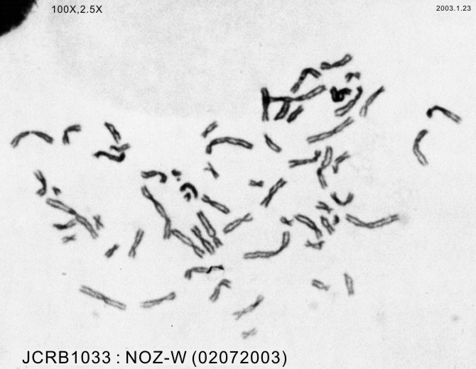
| 遺伝的性質 | No. of chromosome = 46. | 細胞寿命 | infinite |
| クライシスPDL | 形態 | epithelial-like | |
| 一般性状 | Dt=48 hrs. Plating efficiency is 14-19%. Cells are transplantable to nude mice and the morphology of the transplanted tumor was similar to the original one. | 細胞分類 | tumor |
| 細胞樹立者名 | Honma,S. & Nagamori,S. | 細胞寄託者 | Nagamori,S. |
| 分譲時制限 | Require negotiation and agreement from Dr.Nagamori before shipping. | コメント | Cell Bank will handle to get the approval but the depositor may contact to the requested person directly. |
| 入手年 | 2001 | 培養培地 | Williams'E medium with 10% fetal calf serum. |
| 継代方法 | Cells are harvested after treatment with 0.25% trypsin and 0.02% EDTA. | 継代時細胞数 | |
| 人種 | Japanese | 炭酸ガス濃度 | 5 % |
| 採取組織名 | ascites | 組織型 | moderately differentiated adenocarcinoma. |
| ウイルスDNA・RNA検出検査 (Detection of virus genome fragment by Real-time PCR) | |||||||||
|---|---|---|---|---|---|---|---|---|---|
| ウイルスDNA 検出検査 |
tested | ウイルスRNA 検出検査 |
tested | ||||||
| CMV | - | parvoB19 | - | HCV | - | HTLV-1 | - | ||
| EBV | - | HBV | - | HIV-1 | - | HTLV-2 | - | ||
| HHV6 | - | HTLV-1 | - | HIV-2 | - | HAV | - | ||
| HHV7 | - | HTLV-2 | - |
-/negative. +/positive. nt/not tested. (positive (+) does not immediately mean the production of infectious viral particles.) |
|||||
| BKV | - | HIV-1 | - | ||||||
| JCV | - | HIV-2 | - | ||||||
| ADV | - | HPV18 | - | ||||||
| Notes | |||||||||
| Reference | |
|---|---|
| Pubmed id:2543327 | [Combination therapy of hyperthermia and other methods in liver and bile tract cancers--evaluation of these methods using cancer cell lines in vitro]. Hasumura S,Nagamori S,Fujise K,Homma S,Sujino H,Matsuura T,Shimizu K,Niiya M,Kameda H Gan To Kagaku Ryoho. 1989 Apr;16(4 Pt 2-3):1905-12 |
| Research results by users. Click! | |
|---|---|
| Pubmed id:25154789 |
Tumor necrosis factor-α promotes the lymphangiogenesis of gallbladder carcinoma through nuclear factor-κB-mediated upregulation of vascular endothelial growth factor-C. Du Q,Jiang L,Wang X,Wang M,She F,Chen Y Cancer Sci. 2014 Oct;105(10):1261-71 |
| Pubmed id:25165862 |
Schisandrin B induces apoptosis and cell cycle arrest of gallbladder cancer cells. Xiang SS,Wang XA,Li HF,Shu YJ,Bao RF,Zhang F,Cao Y,Ye YY,Weng H,Wu WG,Mu JS,Wu XS,Li ML,Hu YP,Jiang L,Tan ZJ,Lu W,Liu F,Liu YB Molecules. 2014 Aug 27;19(9):13235-50 |
| Pubmed id:25090123 |
Cordycepin induces S phase arrest and apoptosis in human gallbladder cancer cells. Wang XA,Xiang SS,Li HF,Wu XS,Li ML,Shu YJ,Zhang F,Cao Y,Ye YY,Bao RF,Weng H,Wu WG,Mu JS,Hu YP,Jiang L,Tan ZJ,Lu W,Wang P,Liu YB Molecules. 2014 Jul 31;19(8):11350-65 |
| Pubmed id:24756644 |
UHRF1 depletion suppresses growth of gallbladder cancer cells through induction of apoptosis and cell cycle arrest. Qin Y,Wang J,Gong W,Zhang M,Tang Z,Zhang J,Quan Z Oncol Rep. 2014 Jun;31(6):2635-43 |
| Pubmed id:24329682 |
Inhibitory effects of deleted in liver cancer 1 gene on gallbladder cancer growth through induction of cell cycle arrest and apoptosis. Qin Y,Chu B,Gong W,Wang J,Tang Z,Shen J,Quan Z J Gastroenterol Hepatol. 2014 May;29(5):964-72 |
| Pubmed id:24655726 |
Oridonin induces apoptosis and cell cycle arrest of gallbladder cancer cells via the mitochondrial pathway. Bao R,Shu Y,Wu X,Weng H,Ding Q,Cao Y,Li M,Mu J,Wu W,Ding Q,Tan Z,Liu T,Jiang L,Hu Y,Gu J,Liu Y BMC Cancer. 2014 Mar 21;14():217 |
| Pubmed id:24966896 |
miR-1 and miR-145 act as tumor suppressor microRNAs in gallbladder cancer. Letelier P,García P,Leal P,Álvarez H,Ili C,López J,Castillo J,Brebi P,Roa JC Int J Clin Exp Pathol. 2014;7(5):1849-67 |
| Pubmed id:25010679 |
Forkhead box L1 is frequently downregulated in gallbladder cancer and inhibits cell growth through apoptosis induction by mitochondrial dysfunction. Qin Y,Gong W,Zhang M,Wang J,Tang Z,Quan Z PLoS One. 2014;9(7):e102084 |
| Pubmed id:25277242 |
Expression of the RIP-1 gene and its role in growth and invasion of human gallbladder carcinoma. Zhu G,Chen X,Wang X,Li X,Du Q,Hong H,Tang N,She F,Chen Y Cell Physiol Biochem. 2014;34(4):1152-65 |
| Pubmed id:24568162 |
Baicalin induces apoptosis of gallbladder carcinoma cells in vitro via a mitochondrial-mediated pathway and suppresses tumor growth in vivo. Shu YJ,Bao RF,Wu XS,Weng H,Ding Q,Cao Y,Li ML,Mu JS,Wu WG,Ding QC,Liu TY,Jiang L,Hu YP,Tan ZJ,Wang P,Liu YB Anticancer Agents Med Chem. 2014;14(8):1136-45 |
| Pubmed id:23830367 |
Combination chemotherapy of nafamostat mesylate with gemcitabine for gallbladder cancer targeting nuclear factor-κB activation. Iwase R,Haruki K,Fujiwara Y,Furukawa K,Shiba H,Uwagawa T,Misawa T,Ohashi T,Yanaga K J Surg Res. 2013 Sep;184(1):605-12 |
| Pubmed id:22835954 |
Synergic effect of photodynamic therapy using talaporfin sodium with conventional anticancer chemotherapy for the treatment of bile duct carcinoma. Nonaka Y,Nanashima A,Nonaka T,Uehara M,Isomoto H,Abo T,Nagayasu T J Surg Res. 2013 May;181(2):234-41 |
| Pubmed id:24217644 |
Role of aquaporin-5 in gallbladder carcinoma. Sekine S,Shimada Y,Nagata T,Sawada S,Yoshioka I,Matsui K,Moriyama M,Omura T,Osawa S,Shibuya K,Hashimoto I,Watanabe T,Hojo S,Hori R,Okumura T,Yoshida T,Tsukada K Eur Surg Res. 2013;51(3-4):108-17 |
| Pubmed id:22843894 |
Establishment and characterization of a new human gallbladder carcinoma cell line. Sekine S,Shimada Y,Nagata T,Moriyama M,Omura T,Yoshioka I,Hori R,Matsui K,Sawada S,Okumura T,Yoshida T,Tsukada K Anticancer Res. 2012 Aug;32(8):3211-8 |
| Pubmed id:22320835 |
Adenovirus-mediated gene transfer of tissue factor pathway inhibitor-2 inhibits gallbladder carcinoma growth in vitro and in vivo. Qin Y,Zhang S,Gong W,Li J,Jia J,Quan Z Cancer Sci. 2012 Apr;103(4):723-30 |
| Pubmed id:22071224 |
Vascular endothelial growth factor-D promotes growth, lymphangiogenesis and lymphatic metastasis in gallbladder cancer. Lin W,Jiang L,Chen Y,She F,Han S,Zhu J,Zhou L,Tang N,Wang X,Li X Cancer Lett. 2012 Jan 28;314(2):127-36 |
| Pubmed id:22201822 |
SNCG gene silencing in gallbladder cancer cells inhibits key tumorigenic activities. Han S,She F,Wang D,Yao X,Jiang L,Chen Y Front Biosci (Landmark Ed). 2012 Jan 1;17():1589-98 |
| Pubmed id:21360081 |
Advantages of laserphyrin compared with photofrin in photodynamic therapy for bile duct carcinoma. Nonaka T,Nanashima A,Nonaka M,Uehara M,Isomoto H,Nonaka Y,Nagayasu T J Hepatobiliary Pancreat Sci. 2011 Jul;18(4):592-600 |
| Pubmed id:20734221 |
Vascular endothelial growth factor-C promotes the growth and invasion of gallbladder cancer via an autocrine mechanism. Chen Y,Jiang L,She F,Tang N,Wang X,Li X,Han S,Zhu J Mol Cell Biochem. 2010 Dec;345(1-2):77-89 |
| Pubmed id:20651359 |
Analysis of apoptotic effects induced by photodynamic therapy in a human biliary cancer cell line. Nonaka T,Nanashima A,Nonaka M,Uehara M,Isomoto H,Asahina I,Nagayasu T Anticancer Res. 2010 Jun;30(6):2113-8 |
| Pubmed id:20464561 |
Novel photodynamic therapy against biliary tract carcinoma using mono-L: -aspartyl chlorine e6: basic evaluation for its feasibility and efficacy. Kasuya K,Shimazu M,Suzuki M,Kuroiwa Y,Usuda J,Itoi T,Tsuchida A,Aoki T J Hepatobiliary Pancreat Sci. 2010 May;17(3):313-21 |
| Pubmed id:19209029 |
Schedule-dependent synergistic interaction between gemcitabine and oxaliplatin in human gallbladder adenocarcinoma cell lines. Makiyama A,Qin B,Uchino K,Shibata Y,Arita S,Isobe T,Hirano G,Kusaba H,Baba E,Akashi K,Nakano S Anticancer Drugs. 2009 Feb;20(2):123-30 |
| Pubmed id:18204862 |
Upregulation of topoisomerase IIalpha expression in advanced gallbladder carcinoma: a potential chemotherapeutic target. Washiro M,Ohtsuka M,Kimura F,Shimizu H,Yoshidome H,Sugimoto T,Seki N,Miyazaki M J Cancer Res Clin Oncol. 2008 Jul;134(7):793-801 |
| Pubmed id:17235714 |
Antitumor effect of gemcitabine on orthotopically inoculated human gallbladder cancer cells in nude mice. Mita Y,Ajiki T,Kamigaki T,Okazaki T,Hori H,Horiuchi H,Hirata K,Fujita T,Fujimori T,Kuroda Y Ann Surg Oncol. 2007 Apr;14(4):1374-80 |
| Pubmed id:15855925 |
Oncolytic adenoviral therapy in gallbladder carcinoma. Tekant Y,Davydova J,Ramirez PJ,Curiel DT,Vickers SM,Yamamoto M Surgery. 2005 May;137(5):527-35 |
| Pubmed id:15743030 |
A MEK inhibitor (U0126) markedly inhibits direct liver invasion of orthotopically inoculated human gallbladder cancer cells in nude mice. Horiuchi H,Kawamata H,Furihata T,Omotehara F,Hori H,Shinagawa Y,Ohkura Y,Tachibana M,Yamazaki T,Ajiki T,Kuroda Y,Fujimori T J Exp Clin Cancer Res. 2004 Dec;23(4):599-606 |
| Pubmed id:12963974 |
A MEK inhibitor (U0126) prolongs survival in nude mice bearing human gallbladder cancer cells with K-ras mutation: analysis in a novel orthotopic inoculation model. Horiuchi H,Kawamata H,Fujimori T,Kuroda Y Int J Oncol. 2003 Oct;23(4):957-63 |
| Pubmed id:2543327 |
[Combination therapy of hyperthermia and other methods in liver and bile tract cancers--evaluation of these methods using cancer cell lines in vitro]. Hasumura S,Nagamori S,Fujise K,Homma S,Sujino H,Matsuura T,Shimizu K,Niiya M,Kameda H Gan To Kagaku Ryoho. 1989 Apr;16(4 Pt 2-3):1905-12 |
| Pubmed id:3154020 |
[Establishment and characterization of a human gall bladder carcinoma cell line NOZ]. Homma S,Hasumura S,Nagamori S,Kameda H Hum Cell. 1988 Mar;1(1):95-7 |
| Pubmed id:none |
バイオテクノロジー素材としてのヒト癌細胞 (1)。胆嚢癌NOZ。蛋白質。核酸 永森静志 蛋白質・核酸・酵素 33, 2605-2606 (1988) |
| Pubmed id:none |
Morphological changes in human gall bladder carcinoma cell line-NOZ due to epirubicin and doxorubicin Masaaki Kawada, Hideki Aizaki, Kenichi Fukaya, Minoru Niiya, Tomokazu Matsuura, Hajime Sujino, Satoshi Hasumura, Seishi Nagamori, Gotaro Toda Medical Electron Microscopy 27, 175-177 (1994) |
| Pubmed id:none |
閉塞性黄疸に対するPTCD胆汁細胞診とK-ras点突然変異の解析 味木 徹夫, 藤盛 孝博, 塚本 龍子, 渥美 亜紀子, 小野山 裕彦, 前田 盛, 斎藤 洋一 日本臨床細胞学会雑誌 34(3), 424-428 (1995) |
| Pubmed id:none |
微小管を標的とした抗癌剤のヒト胆嚢癌細胞に対する効果に関する基礎的研究 柴本 由香, 高木 一郎 胆道 14(1), 42-50 (2000) |
| Images |
|---|
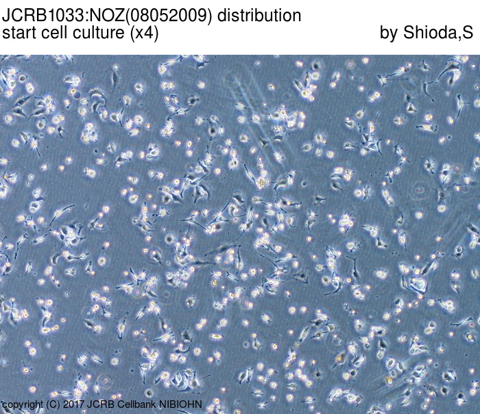 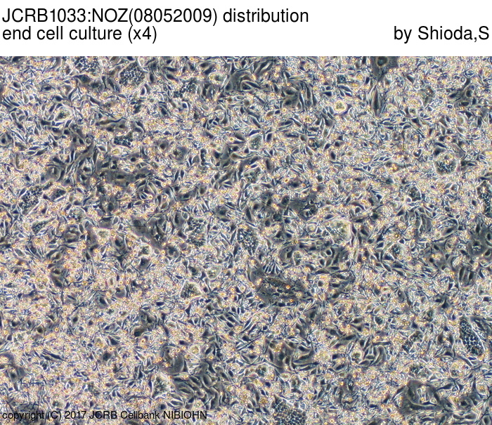 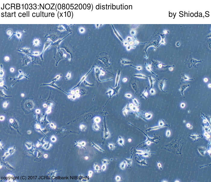 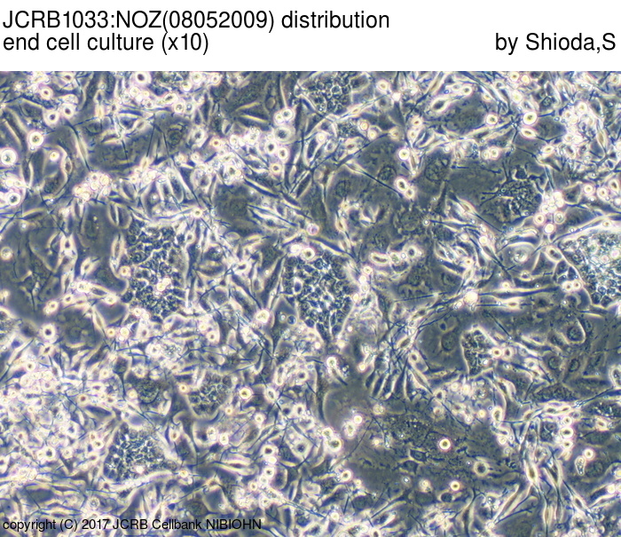     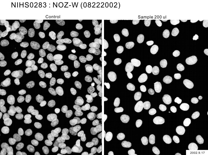                 |
LOT Information
| 細胞番号 | JCRB1033 | 細胞名 | NOZ |
|---|---|---|---|
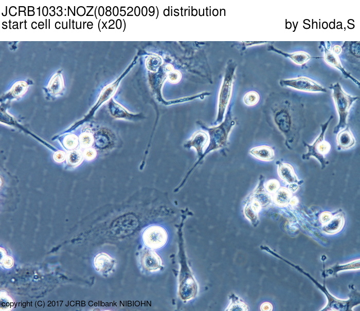
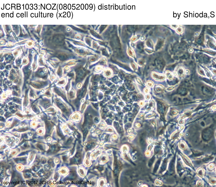
| 培養ロット番号 | 08052009 | 培養種別 | distribution |
| 培地 | William's E medium with10% heat inactivated fetal bovine serum(Intergen RB52305). | 培養温度 | 37 C |
| 継代時細胞数(濃度) | 2.8x10^4 cells/sq.cm | 継代方法 | Cells were harvested after treatment with 0.25%(GIBCO) trypsin and 0.02% EDTA. |
| 増殖速度 | NT | 凍結時生細胞濃度 | 4.5x10^6 |
| 凍結時生細胞率 | 99.1 | 使用抗生物質 | free |
| 継代数 | p7* | PDL数(プライマリ) | NT |
| マイコプラズマ検出 | - | 細菌汚染検出 | - |
| 真菌汚染検出 | - | アイソザイム検査・動物名 | NT |
| 染色体モード | NT | 染色体情報 | NT |
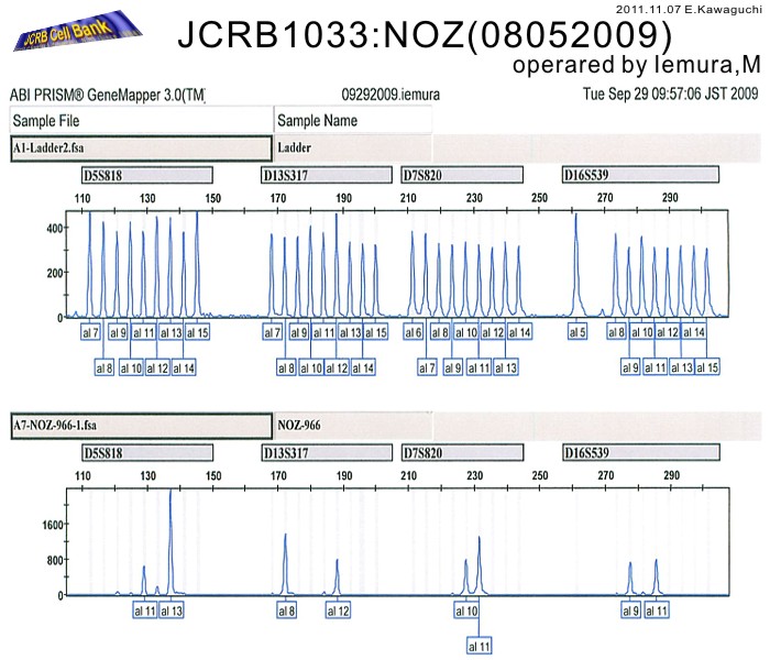
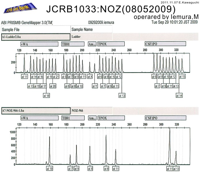
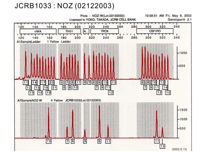
| 表面抗原 | NT | DNA Profile (STR) | D5S818:11,13 D13S317:8,12 D7S820:10,11 D16S539:9,11 VWA:19 TH01:7,9 AM:X TPOX:8,11 CSF1PO:11,13 |
| 接着性 | Yes | 導入外部遺伝子 | NT |
| 凍結培地 | Cell Banker BLC-1(Nihon Zenyaku Industries) | 炭酸ガス濃度 | 5 % |
| 解凍後生細胞率 | 追加情報 |
| Images |
|---|
|
|
| 細胞番号 | JCRB1033 | 細胞名 | |
|---|---|---|---|
| 培養ロット番号 | 09162008 | 培養種別 | distribution |
| 培地 | William's medium E with 10% fetal bovine serum (GIBCO Cat. # 10099, lot 484375) | 培養温度 | 37 C |
| 継代時細胞数(濃度) | 1 - 2 x 10^5 cells/mL | 継代方法 | 0.25% trypsin and 0.02% EDTA |
| 増殖速度 | 30 hrs | 凍結時生細胞濃度 | 4.8 x 10^6 |
| 凍結時生細胞率 | 97.5 | 使用抗生物質 | free |
| 継代数 | Unknown (8 at bank) | PDL数(プライマリ) | |
| マイコプラズマ検出 | - | 細菌汚染検出 | - |
| 真菌汚染検出 | - | アイソザイム検査・動物名 | Confirmed as human by NP, G6PD (type B), MD. |
| 染色体モード | 染色体情報 | ||
| 表面抗原 | DNA Profile (STR) | ||
| 接着性 | Yes | 導入外部遺伝子 | |
| 凍結培地 | 10% DMSO, 20% FBS - William's medium E | 炭酸ガス濃度 | 5% |
| 解凍後生細胞率 | 追加情報 |
| 細胞番号 | JCRB1033 | 細胞名 | NOZ |
|---|---|---|---|
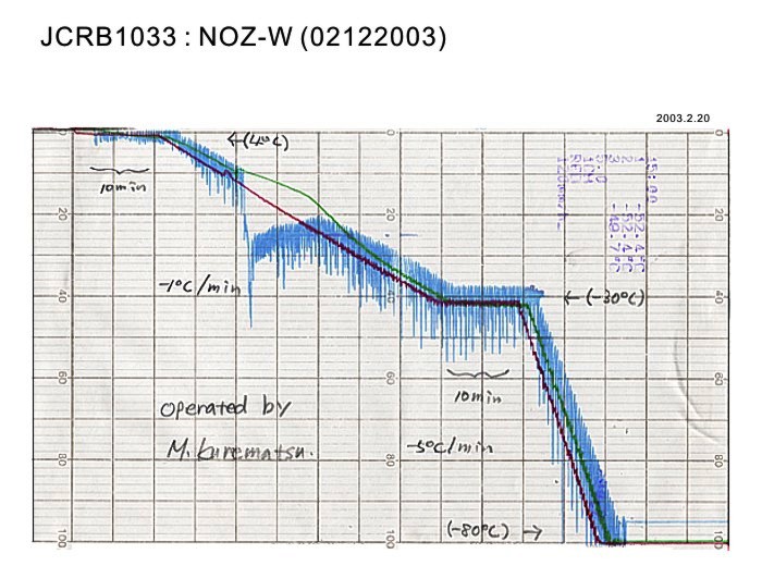
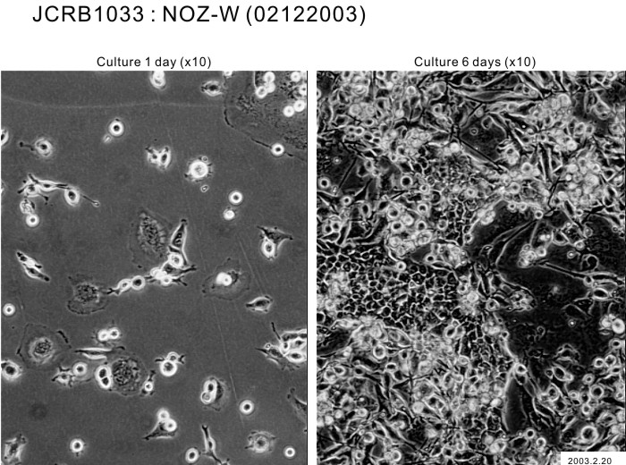
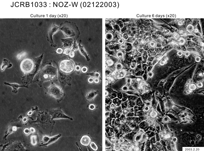
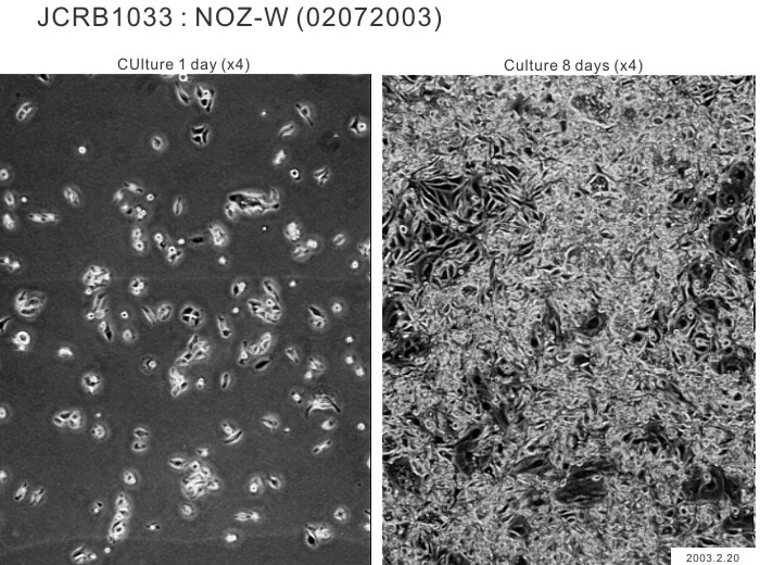
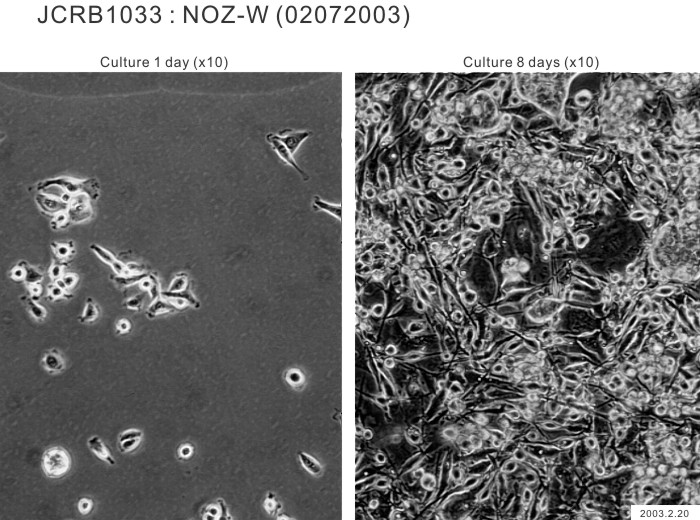
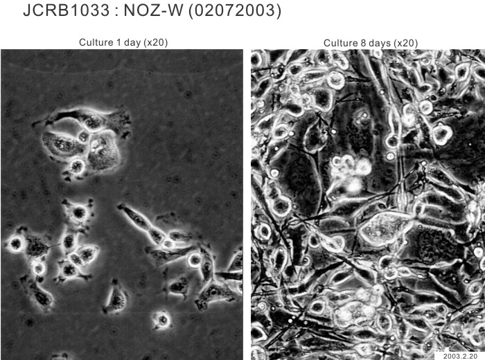
| 培養ロット番号 | 02122003 | 培養種別 | distribution |
| 培地 | William's E medium with 10% fetal calf serum(Intergen RB52305). | 培養温度 | 37 C |
| 継代時細胞数(濃度) | 2.0 x 10^4 cells/sq.cm. | 継代方法 | Cells were harvested after treatment with 0.2% trypsin and 0.02% EDTA. Medium change twice a week and subculture once a week. |
| 増殖速度 | NT | 凍結時生細胞濃度 | 4.5 x 10^6 |
| 凍結時生細胞率 | 98.7 | 使用抗生物質 | free |
| 継代数 | P5* | PDL数(プライマリ) | NT |
| マイコプラズマ検出 | - | 細菌汚染検出 | - |
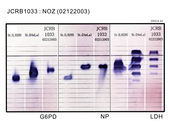
| 真菌汚染検出 | - | アイソザイム検査・動物名 | G6PD(typeB), NP, LDH examined. Human, not HeLa. |
| 染色体モード | NT | 染色体情報 | NT |
| 表面抗原 | NT | DNA Profile (STR) | D5S818:11,13 D13S317:8,12 D7S820:10,11 D16S539:9,11 VWA:19 TH01:7,9 AM:X TPOX:8,11 CSF1PO:11,13 |
| 接着性 | Yes | 導入外部遺伝子 | NT |
| 凍結培地 | Culture medium containing 5% DMSO. | 炭酸ガス濃度 | 5 % |
| 解凍後生細胞率 | 追加情報 |
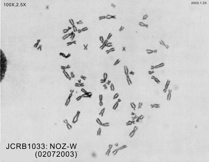
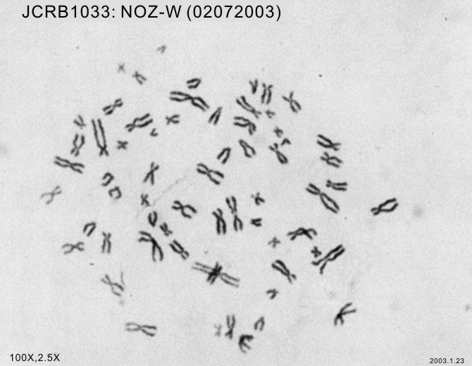
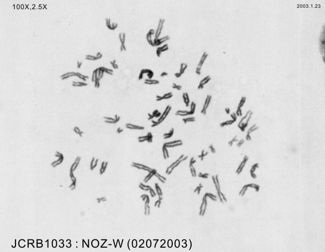
| Images |
|---|
    |
| 細胞番号 | JCRB1033 | 細胞名 | NOZ |
|---|---|---|---|
| 培養ロット番号 | 05192020 | 培養種別 | distribution |
| 培地 | William's medium E with 10% fetal bovine serum (FBS; Sigma Cat. # 172012) | 培養温度 | 37 C |
| 継代時細胞数(濃度) | 6.2 x 10^4 cells/mL | 継代方法 | Cells were harvested after treatment with 0.2% trypsin and 0.02% EDTA. |
| 増殖速度 | approx. 27 hrs. | 凍結時生細胞濃度 | 83 |
| 凍結時生細胞率 | 4.5 x 10^6 | 使用抗生物質 | free |
| 継代数 | Unknown (7 at bank) | PDL数(プライマリ) | |
| マイコプラズマ検出 | - | 細菌汚染検出 | - |
| 真菌汚染検出 | - | アイソザイム検査・動物名 | |
| 染色体モード | 染色体情報 | ||
| 表面抗原 | DNA Profile (STR) | ||
| 接着性 | Yes | 導入外部遺伝子 | |
| 凍結培地 | 炭酸ガス濃度 | 5% | |
| 解凍後生細胞率 | 76 | 追加情報 |